HUMAN SKELETAL GENERAL FEATURES
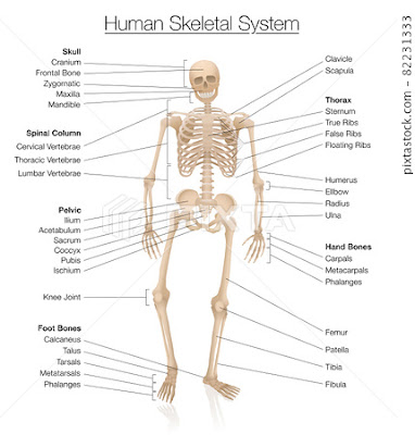
The skeletal system is your body's central framework. The human skeletal system provides support and protection for the body's organs and tissues, as well as allowing for movement. It is composed of bones, cartilage, and ligaments, and can be divided into two main parts: the axial skeleton and the appendicular skeleton.
- The axial skeleton includes the skull, vertebral column, and rib cage, and serves to protect the brain, spinal cord, and organs of the thorax.
- The appendicular skeleton includes the bones of the arms, legs, pelvis, and shoulder girdle, and allows for movement and locomotion.
👉Some general features of the human skeletal system include:
💪Bones are classified according to their shape, which can be long, short, flat, or irregular.
💪Bones are composed of dense, mineralized tissue called cortical bone, and porous, spongy tissue called cancellous bone.
💪Bones are connected to each other by joints, which can be movable or immovable, depending on their location and function.
💪Cartilage is a flexible connective tissue that covers the ends of bones where they meet to form joints.
💪Ligaments are strong, fibrous bands of connective tissue that connect bones to other bones, providing stability and support to the joints.
💪The skeletal system is constantly undergoing remodeling, with bone tissue being broken down and rebuilt throughout life.
💪The bones of the human body are constantly growing and changing throughout life, with peak bone mass typically reached in early adulthood.
Overall, the human skeletal system is an intricate and dynamic system that plays a vital role in the functioning of the human body.
SKULL
The skull is a complex structure composed of several bones that form the head and protect the brain. It consists of two main parts: the cranial bones, which enclose the brain, and the facial bones, which form the front part of the skull.
The cranial bones include the frontal bone, two parietal bones, two temporal bones, the occipital bone, the sphenoid bone, and the ethmoid bone. These bones are flat and provide a protective enclosure for the brain. The skull also contains a number of foramina, or openings, which allow nerves, blood vessels, and other structures to pass through.
The facial bones include the maxilla, mandible, zygomatic bone, nasal bone, lacrimal bone, palatine bone, and inferior nasal concha. These bones give the face its shape and provide attachment points for the muscles involved in chewing, speaking, and making facial expressions.
The skull also contains several important structures, including the cranial vault, which protects the brain, the cranial base, which provides support for the brain and connects the skull to the spinal column, and the facial skeleton, which protects the eyes, nose, and mouth.
In addition to its protective functions, the skull also serves as an attachment point for many muscles involved in head and neck movement. It also contains several openings for the passage of nerves and blood vessels and houses several important sensory organs, including the eyes, ears, and nose.
Overall, the skull is a complex and highly specialized structure that plays a vital role in protecting the brain and supporting the functions of the head and neck.
HUMERUS
The humerus is the long bone located in the upper arm, between the shoulder and elbow joints. It is the largest bone in the upper limb and is essential for movements of the arm, including lifting, pushing, and pulling.
The humerus has a number of distinguishing features that are important for its function. Here are some of the key details of the humerus:
👉Anatomy: The humerus has a shaft or diaphysis, which is the long, tubular portion of the bone that makes up most of its length. At either end of the shaft, there are two distinct epiphyses, which are the rounded ends of the bone that articulate with other bones in the shoulder and elbow joints. The upper epiphysis of the humerus is called the humeral head, while the lower epiphysis is divided into the capitulum, trochlea, and medial and lateral epicondyles.
👉Articulation: The humeral head articulates with the glenoid fossa of the scapula (shoulder blade) to form the glenohumeral joint, which is the main joint of the shoulder. The capitulum articulates with the head of the radius to form the radiocapitellar joint, while the trochlea articulates with the ulna to form the humeroulnar joint. The lateral and medial epicondyles are attachment sites for muscles and ligaments that help stabilize the elbow joint.
👉Blood supply: The humerus is supplied by several arteries, including the humeral circumflex artery, the anterior humeral circumflex artery, and the posterior humeral circumflex artery. These arteries provide blood to the bone and surrounding tissues, helping to nourish and oxygenate them.
👉Muscles: The humerus serves as an attachment site for several important muscles, including the deltoid, rotator cuff, biceps brachii, triceps brachii, and brachialis muscles. These muscles help to control the movement of the shoulder and elbow joints.
👉Fractures: Fractures of the humerus can occur due to trauma or overuse injuries, such as repetitive stress from throwing or weightlifting. Treatment options for humeral fractures may include immobilization with a cast or brace, physical therapy, or surgery in more severe cases.
Overall, the humerus is a critical bone in the upper arm that plays an essential role in the movement and stability of the shoulder and elbow joints.
RADIUS & ULNA
The radius and ulna are two bones located in the forearm, between the elbow and wrist joints. They are parallel to each other and work together to allow movements of the forearm and hand. Here are some key details about the radius and ulna:
👉Anatomy: The radius is the shorter and thicker of the two bones and is located on the thumb side of the forearm. The ulna is longer and thinner and is located on the pinky side of the forearm. The proximal end of the radius articulates with the capitulum of the humerus, while the distal end articulates with the carpal bones in the wrist. The proximal end of the ulna articulates with the trochlea of the humerus and the radius, while the distal end articulates with the radius and the bones of the wrist.
👉Movement: The radius and ulna work together to allow movements of the forearm, including pronation (turning the palm down) and supination (turning the palm up). These movements are facilitated by the rotation of the radius around the ulna.
Muscles: The radius and ulna serve as attachment sites for several important muscles that control movements of the forearm and hand, including the flexor and extensor muscles of the wrist and fingers.
👉Fractures: Fractures of the radius and ulna can occur due to trauma or overuse injuries, such as repetitive stress from activities like tennis or typing. Treatment options for fractures may include immobilization with a cast or brace, physical therapy, or surgery in more severe cases.
Overall, the radius and ulna are two important bones in the forearm that work together to allow movements of the forearm and hand. They play a crucial role in activities of daily living and in many sports and recreational activities.
FEMUR
The femur, also known as the thigh bone, is the longest, largest, and strongest bone in the human body. It is located in the thigh region and connects the hip joint to the knee joint. Here are some key details about the femur:
👉Anatomy: The femur has a shaft or diaphysis, which is the long, tubular portion of the bone that makes up most of its length. At either end of the shaft, there are two distinct epiphyses, which are the rounded ends of the bone that articulate with other bones in the hip and knee joints. The upper epiphysis of the femur is called the femoral head, while the lower epiphysis is divided into the medial and lateral condyles and the patellar groove.
👉Articulation: The femoral head articulates with the acetabulum of the pelvis to form the hip joint, while the medial and lateral condyles articulate with the tibia to form the knee joint. The patellar groove provides a space for the patella (kneecap) to glide during knee movements.
👉Blood supply: The femur is supplied by several arteries, including the medial and lateral femoral circumflex arteries and the deep femoral artery. These arteries provide blood to the bone and surrounding tissues, helping to nourish and oxygenate them.
👉Muscles: The femur serves as an attachment site for several important muscles, including the quadriceps, hamstrings, and gluteal muscles. These muscles help to control the movement of the hip and knee joints.
👉Fractures: Fractures of the femur can occur due to trauma or overuse injuries, such as repetitive stress from running or jumping. Treatment options for femoral fractures may include immobilization with a cast or brace, physical therapy, or surgery in more severe cases.
Overall, the femur is a critical bone in the human body that plays an essential role in the movement and stability of the hip and knee joints. It supports the weight of the body and allows for a wide range of movements, including walking, running, jumping, and climbing.
TIBIA & FIBULA
The tibia and fibula are two bones located in the lower leg between the knee and ankle joints. They work together to provide support, stability, and movement to the lower leg and foot. Here are some key details about the tibia and fibula:
👉Anatomy: The tibia, also known as the shinbone, is the larger and stronger of the two bones and is located on the medial (inner) side of the lower leg. The fibula is smaller and more slender and is located on the lateral (outer) side of the lower leg. The proximal end of the tibia articulates with the femur to form the knee joint, while the distal end articulates with the ankle bones. The fibula does not directly articulate with the knee joint, but it does contribute to the ankle joint.
👉Movement: The tibia and fibula work together to allow movements of the lower leg and foot, including dorsiflexion (lifting the foot up) and plantarflexion (pointing the foot down).
👉Muscles: The tibia and fibula serve as attachment sites for several important muscles that control movements of the lower leg and foot, including the calf muscles, ankle dorsiflexion, and ankle evertors.
👉Fractures: Fractures of the tibia and fibula can occur due to trauma or overuse injuries, such as repetitive stress from activities like running or jumping. Treatment options for fractures may include immobilization with a cast or brace, physical therapy, or surgery in more severe cases.
Overall, the tibia and fibula are two important bones in the lower leg that work together to provide support, stability, and movement to the lower leg and foot. They play a crucial role in activities of daily living and in many sports and recreational activities.
VERTEBRA
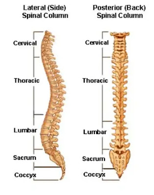
There are 33 vertebrae in the human spine, divided into five regions: cervical, thoracic, lumbar, sacral, and coccygeal. Each region has a slightly different shape and function, as described below:
👉Cervical vertebrae: There are seven cervical vertebrae in the neck region, numbered C1 to C7. They are the smallest vertebrae and are unique in having transverse foramen on each side of the vertebral body, through which the vertebral arteries pass to supply blood to the brain. The first cervical vertebra called the atlas, supports the skull, while the second cervical vertebra, called the axis, allows for rotation of the head.
👉Thoracic vertebrae: There are twelve thoracic vertebrae in the upper back region, numbered T1 to T12. They are larger than the cervical vertebrae and have long, curved spinous processes that form the prominent bumps on the back. The thoracic vertebrae also have facets for articulation with the ribs, forming the thoracic cage that protects the heart and lungs.
👉Lumbar vertebrae: There are five lumbar vertebrae in the lower back region, numbered L1 to L5. They are the largest and strongest of the vertebrae, with thick, block-like bodies and short, stumpy spinous processes. The lumbar vertebrae bear the most weight and are prone to degenerative changes and injury.
👉Sacral vertebrae: The sacrum is a single bone formed by the fusion of five sacral vertebrae, located at the base of the spine. They are triangular in shape and form the back wall of the pelvic cavity, providing support and attachment for the pelvic organs and lower limbs.
👉Coccygeal vertebrae: The coccyx, or tailbone, is a single bone formed by the fusion of four coccygeal vertebrae, located at the bottom of the spine. They are small and rudimentary, with no distinct features or functions.
Overall, each type of vertebra has a unique shape and function that contributes to the overall structure and function of the spine. Together, they provide support and protection for the spinal cord, allow for movement and flexibility, and play a crucial role in overall body posture and alignment.
SCAPULA
The scapula, also known as the shoulder blade, is a flat, triangular-shaped bone located on the upper back. It plays a crucial role in the movement and stability of the shoulder joint, as well as the overall posture and alignment of the upper body. Here are some key details about the scapula:
👉Anatomy: The scapula is located on the posterior (back) aspect of the thorax, and it connects to the clavicle (collarbone) and humerus (upper arm bone) to form the shoulder joint. It has three main parts: the body, which is the large, flat triangular portion of the bone; the acromion process, which is the flat, bony projection that forms the top of the shoulder and articulates with the clavicle; and the coracoid process, which is the hook-shaped projection on the front of the scapula that serves as an attachment site for several muscles and ligaments.
👉Muscles: The scapula serves as an attachment site for several important muscles that control movements of the shoulder joint, including the trapezius, serratus anterior, and rotator cuff muscles.
👉Movement: The scapula moves in coordination with the humerus and clavicle to allow a wide range of shoulder movements, including elevation, depression, protraction, retraction, and rotation.
👉Injuries: Injuries to the scapula can occur due to trauma or overuse injuries, such as rotator cuff tears, shoulder dislocations, or fractures. Treatment options for scapula injuries may include immobilization with a sling, physical therapy, or surgery in more severe cases.
Overall, the scapula is an important bone in the upper body that plays a crucial role in the movement and stability of the shoulder joint, as well as the overall posture and alignment of the upper body. It is a key attachment site for several important muscles and is prone to injury due to its position and function.
RIBS
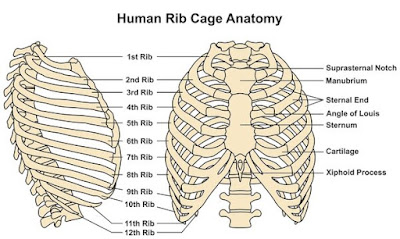
Ribs are long, curved bones that form the thoracic cage, which protects the heart and lungs in the chest cavity. There are 12 pairs of ribs in the human body, and they are numbered from 1 to 12, with the first pair being the shortest and the last pair being the longest. Here are some key details about ribs:
👉Anatomy: Each rib has two ends: the sternal end, which is the anterior end that attaches to the sternum (breastbone), and the vertebral end, which is the posterior end that attaches to the thoracic vertebrae. The upper seven pairs of ribs are called true ribs because they attach directly to the sternum via costal cartilage, while the lower five pairs are called false ribs because they attach indirectly to the sternum or not at all. The last two pairs of ribs, which are the lowest, are also called floating ribs because they do not attach to the sternum at all.
👉Function: The ribs serve several important functions, including protecting the heart and lungs from injury, providing support for the chest wall and upper body, and assisting in the process of breathing. The ribs expand and contract during respiration, helping to increase and decrease the volume of the thoracic cavity and allowing air to be drawn in and expelled from the lungs.
👉Injuries: Rib fractures are a common type of chest injury that can result from trauma, such as a fall or motor vehicle accident, or from repetitive overuse, such as in sports or heavy lifting. Treatment for rib fractures may include pain management, rest, and in some cases, surgery.
👉Variations: Some people may have additional or missing ribs, which can occur due to genetic variations or developmental abnormalities. Extra ribs, known as cervical ribs, can cause compression of nerves or blood vessels in the neck, while missing ribs may not cause any significant problems.
Overall, the ribs play an important role in protecting the heart and lungs, providing support for the chest wall and upper body, and assisting in the process of breathing. Rib injuries can be painful and potentially serious, and may require medical attention depending on the severity and extent of the injury.
PELVIC BONE
The pelvic bone, also known as the hip bone or innominate bone, is a large, irregularly shaped bone that forms the pelvic girdle, which connects the spine to the lower limbs. The pelvic bone consists of three parts: the ilium, ischium, and pubis, which are all separate bones in childhood but fuse together during adolescence to form the adult hip bone. Here are some key details about the pelvic bone:
👉Anatomy: The ilium is the largest and most superior part of the pelvic bone, forming the hip bone's bulk. The ischium forms the posterior and inferior part of the hip bone, while the pubis is the anterior and inferior part of the bone. The three parts of the pelvic bone join together at the acetabulum, which is the socket where the femur (thigh bone) articulates to form the hip joint.
👉Function: The pelvic bone plays a crucial role in supporting the weight of the body and providing attachment sites for muscles that move the hip joint, thigh, and lower back. The pelvic bone also protects the reproductive and urinary organs in the pelvic cavity.
👉Gender differences: The male and female pelvis have some distinct differences due to the needs of childbirth. The female pelvis is wider and shallower, with a larger pelvic outlet, to allow for the passage of a baby during childbirth. The male pelvis is narrower and deeper, with a smaller pelvic outlet.
👉Injuries: Pelvic fractures can occur due to high-energy trauma, such as a car accident or fall from a height, and can be potentially life-threatening due to the proximity of major blood vessels and organs in the pelvic cavity. Treatment for pelvic fractures may include surgery, pain management, and rehabilitation.
Overall, the pelvic bone is a complex structure that plays a crucial role in supporting the weight of the body, providing attachment sites for muscles, and protecting internal organs. Gender differences in the pelvic bone reflect the unique needs of childbirth. Pelvic fractures can be serious and require medical attention.


%20(1).jpeg)
%20(1).jpeg)