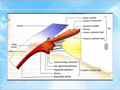
- Ciliary body is forward continuation of the choroid at ora serrata.
- In cut-section, it is triangular in shape. The anterior side of the triangle forms the part of the angle of anterior and posterior chambers. In its middle the iris is attached.
- The outer side of the triangle lies against the sclera with a suprachoroidal space in between. The inner side of the triangle is divided into two parts.
- The anterior part (about 2 mm) having finger-like ciliary processes is called pars plicata and the posterior smooth part (about 4 mm) is called pars plana.
Microscopic structure: From without inwards ciliary body consists of following five layers:
1. Supraciliary lamina. It is the outermost condensed part of the stroma and consists of pigmented collagen fibres. Posteriorly, it is the continuation of suprachoroidal lamina and anteriorly it becomes continuous with the anterior limiting membrane of iris
2. Stroma of the ciliary body. It consists of connective tissue of collagen and fibroblasts. Embedded in the stroma are ciliary muscle, vessels, nerves, pigment and other cells. Ciliary muscle occupies most of the outer part of ciliary body. In cut section it is triangular in shape. It is a non-striated muscle having three parts: (i) the longitudinal or meridional fibres which help in aqueous outflow; (ii) the circular fibres which help in accommodation; and (iii) the radial or oblique fibres act in the same way as the longitudinal fibres. Ciliary muscle is supplied by parasympathetic fibres through the short ciliary nerves.
3. Layer of pigmented epithelium. It is the forward continuation of the retinal pigment epithelium. Anteriorly it is continuous with the anterior pigmented epithelium of the iris.
4. Layer of non-pigmented epithelium. It consists mainly of low columnar or cuboidal cells, and is the forward continuation of the sensory retina. It continues anteriorly as the posterior (internal) pigmented epithelium of the iris.
5. Internal limiting membrane. It is the forward continuation of the internal limiting membrane of the retina. It lines the non-pigmented epithelial layers.
FUNCTIONS OF CILIARY BODY:
- Site of aqueous humour production
- Maintenance of IOP
- Constitutes blood-aqueous barrier
- Ciliary muscle helps in Accommodation
- ciliary muscles help in opening up the schlemn's canal and that helps in aqueous outflow
Pars plicata:The pars plicata is the portion of the ciliary body that contains the ciliary processes. Ciliary processes are the finger-like projections, which extend into the posterior chamber. The regions between ciliary processes are called valleys of Kuhnt. They are approximately 70 to 80 in numbers. A ciliary process measures approximately 2 mm in length, 0.5 mm in width, and 1 mm in height.
Pars plana:As discussed earlier, pars plana is the flat or smooth part of the ciliary body. It terminates at the ora serrata, which is the transitional zone between the ciliary body and choroid. Histologically, the pars plana consist of a double layer of epithelial cells: the inner, nonpigmented epithelium, which is continuous with neurosensory retina; and the outer, pigmented epithelium, which is continuous with the retinal pigment epithelium (RPE).
Ciliary muscle:
3 groups of smooth muscle fibers which function as unit
1.Longitudinal muscle(Outer)- • Attached anteriorly to scleral spur, outer trabecular meshwork
2.Radial muscle/oblique- • Attached to the inner uveal mesh work
3.Circular muscle(inner muscle) • Primarily attached to the ciliary and iris stroma Posteriorly muscle are attached to- elastic structure of par planna vessels and Bruch’ s elastic


%20(1).jpeg)
%20(1).jpeg)